Butterfly wings, fish fins, and human limbs develop precisely and symmetrically. While genetics and chemical environment significantly influence their development, recent research has revealed that mechanical forces play a pivotal role as well.
Piezo proteins have the unique ability to convert mechanical forces—such as the pressure and stretch of developing cells—into chemical signals. While these proteins have been previously shown to regulate blood pressure and sense pain, chemical and biomolecular engineers at the University of Notre Dame have demonstrated their crucial role in organ growth, regulating organ size, and the arrangement of cells in organ tissue.
Their results were published in Cell Reports.
“Piezo acts like the thermostat in your house, it’s constantly measuring and adjusting the conditions of the cells,” said Jeremiah Zartman, associate professor of chemical and biomolecular engineering at the University of Notre Dame.
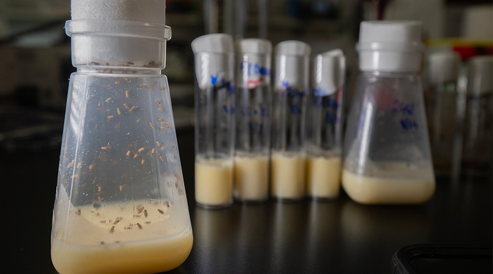
Fruit flies, with their fully sequenced genome, provided Zartman’s lab with a well-studied model for organ development that has many commonalities with organ growth in humans.

While it was known that Piezo proteins play a multifaceted role in cell growth and differentiation, the researchers were after a holistic picture of how this protein functioned on the larger scale of organ development.
On a cellular level, Piezo proteins join to create a gated channel into the cell membrane. Under mechanical tension, the channel’s gate opens, allowing calcium ions to flow in. The concentration of these ions cues the cell’s next step—proliferate, push other cells out, or undergo programmed cell death.
Chemically stimulating Piezo in developing fruit fly wings increases cell division (video courtesy of Zartman Lab)
The team reverse engineered this signaling process, altering the levels of Piezo with drugs or genetic manipulations to better understand how these channels function. The resulting wing asymmetries, aberrant cell death, and changes in cellular proliferation highlighted the protein’s fundamental importance in organ development, even to non-adjacent tissues.
“It was a major surprise to us to find a protein, one that exists predominantly in the cell membrane, that could specifically control robustness, or precision,” said Zartman. “Piezo regulates how cells interact with each other, reach a certain size, stop growing, and it does this to ensure significant precision, there’s very little difference between one side of an organ and the other.”
Moving forward, Zartman said that his multi-institutional team will use mice and fish to explore how Piezo signals healthy cell development versus cancerous growth.


The team’s ongoing work is funded by the National Science Foundation’s (NSF) Emergent Mechanisms in Biology of Robustness, Integration & Organization (EMBRIO) Institute and was supported by the NIH’s National Institute of General Medical Science (NIGMS) with early support from the NSF-Simons Center for Quantitative Biology Pilot program.
— Karla Cruise, Notre Dame Engineering; Photos by Wes Evard, Notre Dame Engineering